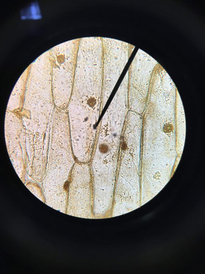
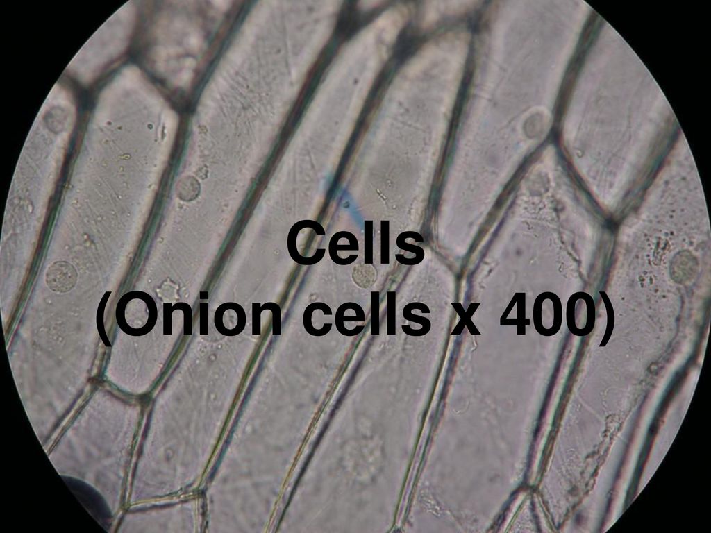
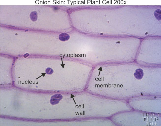
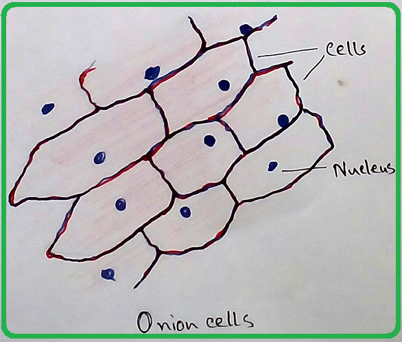
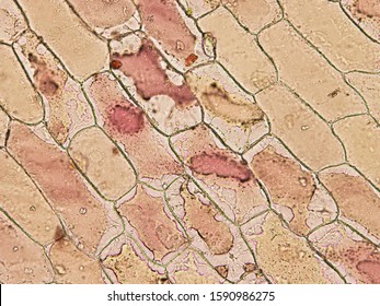
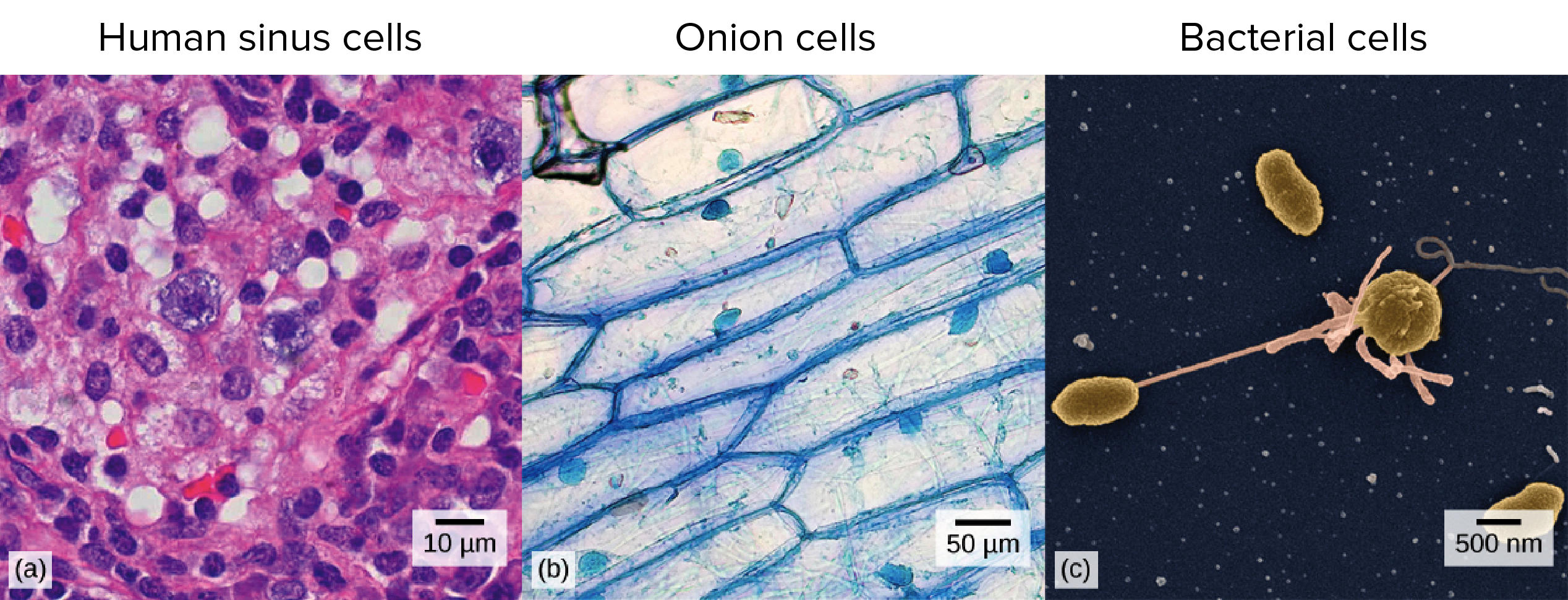
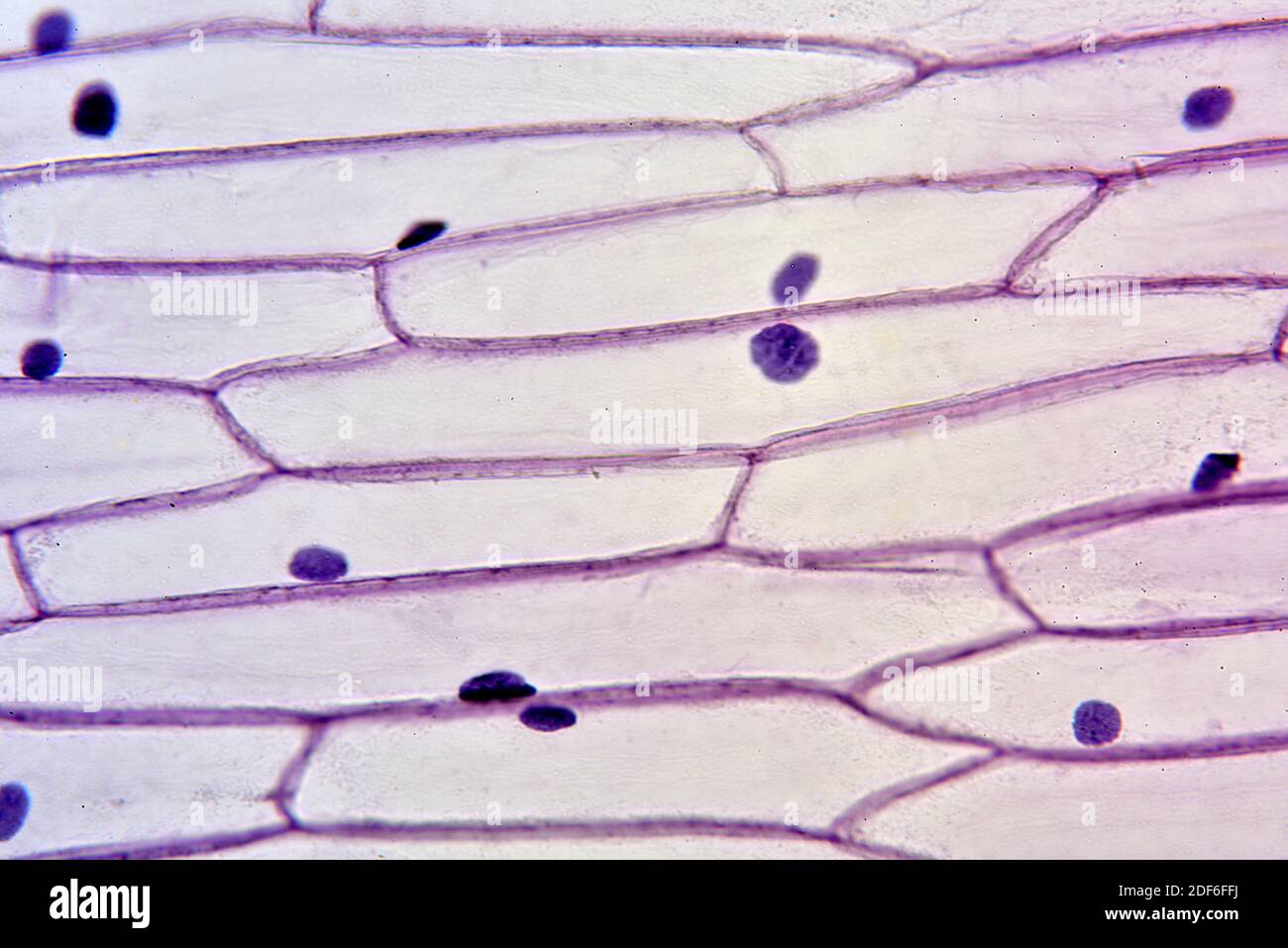
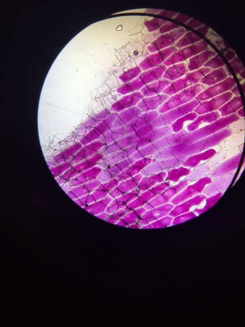
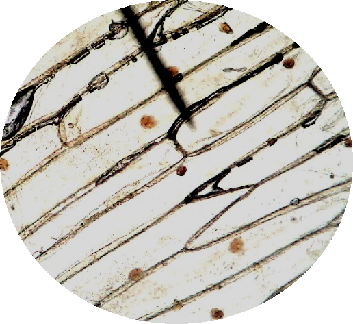
43 onion cells under microscope with labels
Onion Cells Under a Microscope - Requirements/Preparation/Observation Add a drop of iodine solution on the onion membrane (or methylene blue) Gently lay a microscopic cover slip on the membrane and press it down gently using a needle to remove air bubbles. Touch a blotting paper on one side of the slide to drain excess iodine/water solution, Place the slide on the microscope stage under low power to observe. Onion Plant Cell Under Microscope Labeled - Ismael Dauila Explore diffusion/osmosis by looking at onion cells under the microscope. It is used for treating a parasite disease called ich (ichthyophthirius multifiliis; Label the cell wall and chloroplasts. Students will observe plant cells using a light microscope.
Under the Micrsocope: Onion Cell (100x - 400x) - YouTube In this "experiment" we will see onion cells under the microscope.For the experiment you will only need onion, dropper and the microscope (container and tool...

Onion cells under microscope with labels
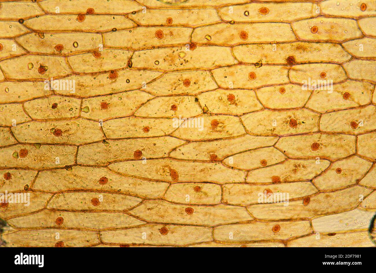
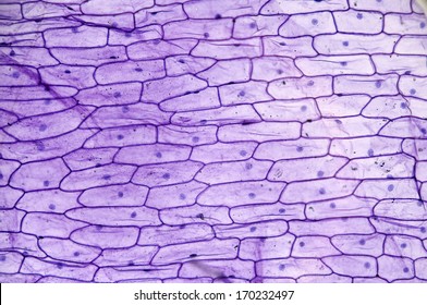
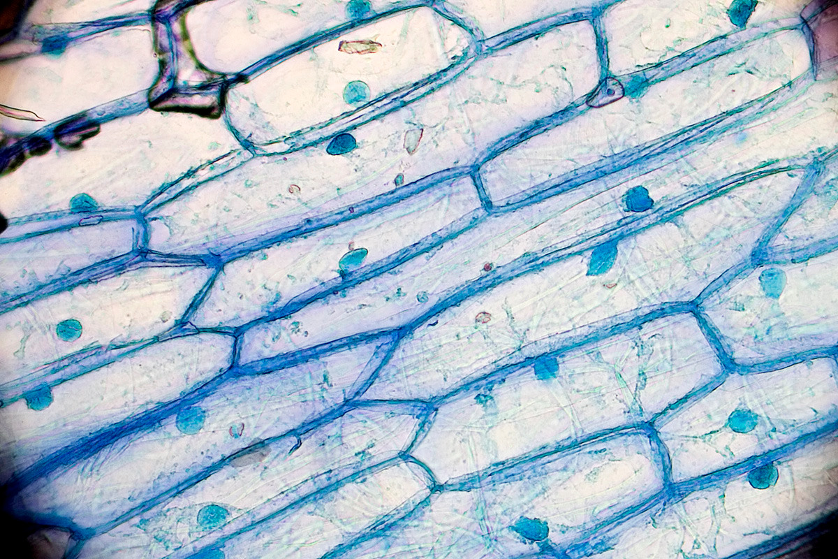
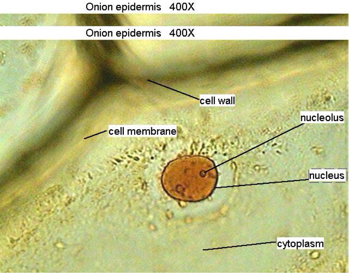
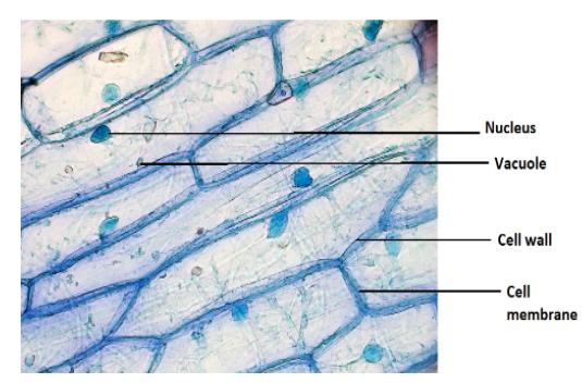
Lesson 3: Onion Dissection & "Look at the Plant Cells" Preparing onion cells slide for a microscope Peel the brown skin away from the outside of the onion. Take one layer of the onion flesh and carefully cut out a piece. On the inside of this piece is a thin sheet of the membrane. Use tweezers or dissection needle to peel the membrane away. What organelles are in an onion cell? - Biology Stack Exchange You cannot see most of these as they appear translucent as well as being too small to see under the light microscope. You need an electron microscope to view these. ... chloroplasts are not present in an onion cell as it is not a photosynthesising cell. This is a typical onion cell slide with labels: Share. Improve this answer. Follow edited ... Onion Cells Stock Photos, Pictures & Royalty-Free Images - iStock Onion epidermis with large cells under microscope Onion epidermis under light microscope. Purple colored, large epidermal cells of an onion, Allium cepa, in a single layer. Each cell with wall, membrane, cytoplasm, nucleus and large vacuole. Photo. Microscope image of plant cells with three nuclei in anaphase
Onion cells under microscope with labels. Scanning electron microscope - Wikipedia History. An account of the early history of scanning electron microscopy has been presented by McMullan. Although Max Knoll produced a photo with a 50 mm object-field-width showing channeling contrast by the use of an electron beam scanner, it was Manfred von Ardenne who in 1937 invented a microscope with high resolution by scanning a very small raster with a demagnified and finely focused ... The Cell Structure of an Onion | Sciencing Onion cells are among the most common choices for cell studies in early biology classes. Easily obtained, inexpensive, they offer samples with no difficult technique required. The thin layer of skin found on the inside of an onion scale (one layer of onion) lifts off without effort and can be wet mounted on a slide with no need for extreme skill. Observing Onion Cells Under The Microscope » Microscope Club Afterwards, carefully mount the prepared and stained onion cell slide onto the microscope stage. Make sure that the cover slip is perfectly aligned with the microscope slide, and that any excess stain has been wiped off. Secure the slide on the stage using the stage clips. IDM Members Meeting Dates 2022 | Institute Of Infectious ... Feb 16, 2022 · IDM Members' meetings for 2022 will be held from 12h45 to 14h30.A zoom link or venue to be sent out before the time.. Wednesday 16 February; Wednesday 11 May; Wednesday 10 August
PPIC Statewide Survey: Californians and Their Government Oct 27, 2022 · Key Findings. California voters have now received their mail ballots, and the November 8 general election has entered its final stage. Amid rising prices and economic uncertainty—as well as deep partisan divisions over social and political issues—Californians are processing a great deal of information to help them choose state constitutional officers and state legislators and to make ... Onion Cell Lab Report.docx - Onion Cell Lab Report By station, remove the single layer of epidermal cells from inner side of the scale leaf. 3(Place the single layer of onion on a glass slide. 4(Place a drop of iodine stain on your onion tissue. 5(Put the cover slip on the stained tissue and gently tap out any air bubbles. 6(Observe the cells under the microscope and see you results. Celery - Wikipedia A celery stalk readily separates into "strings" which are bundles of angular collenchyma cells exterior to the vascular bundles. Wild celery, Apium graveolens var. graveolens, grows to 1 m (3 ft 3 in) tall. Celery is a biennial plant that occurs around the globe. It produces flowers and seeds only during its second year. onion cells under a microscope labeled - sevadham.net onion cells under a microscope labeled. dream team 1992 training. onion cells under a microscope labeledprogramming in scala github. black white and cream living room ideas ...
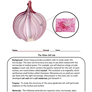
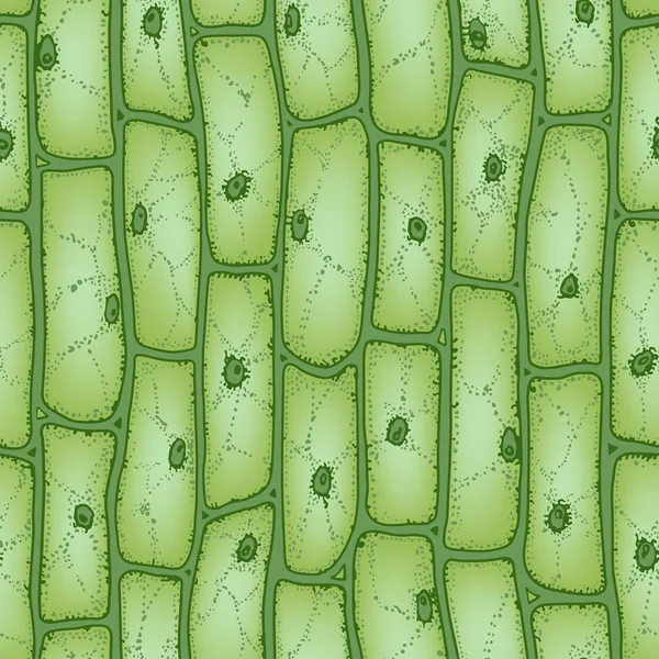
Structure and function of mitochondrial membrane protein ... Oct 29, 2015 · Biological energy conversion in mitochondria is carried out by the membrane protein complexes of the respiratory chain and the mitochondrial ATP synthase in the inner membrane cristae. Recent advances in electron cryomicroscopy have made possible new insights into the structural and functional arrangement of these complexes in the membrane, and how they change with age. This review places ... Onion Cells Under the Microscope LAB - Google Docs.pdf Observations Large, rectangular interlocking cells, Clearly visible distinct cell walls surrounding the cells, Dark stained nucleus, Large vacuoles at the center, Small granules may be observed inside the cells (within the cytoplasm) The layers of an onion contain simple sugars (carbohydrates) some of which are stored as starch (starch granules). Onion peel under microscope | AQuriousMind All living things are made of cells. An onion cell is a multicellular (consisting of many cells) plant organism. An onion cell, like all plant cells, contains the following essential parts: A thick cell wall made of cellulose. Yes, the same thing that makes up your cello tape. It's the cellulose that makes the cell rigid. A cell membrane is ... DOC The Onion Cell Lab - chsd.us Onion tissue provides excellent cells to study under the microscope. The main cell structures are easy to see when viewed with the microscope at medium power. For example, you will observe a large circular . nucleus. in each cell, which contains the genetic material for the cell. In each nucleus, are round bodies called . nucleoli
Lyft's Commitment to Climate Action - Lyft Blog A lot of voters agree with us. Early support for the measure is strong. What started with good policy created by a diverse group of organizations — including the Natural Resources Defense Council, the American Lung Association, California State Firefighters, the Coalition for Clean Air, the State Association of Electrical Workers – IBEW, the San Francisco Bay Area Planning and Urban ...
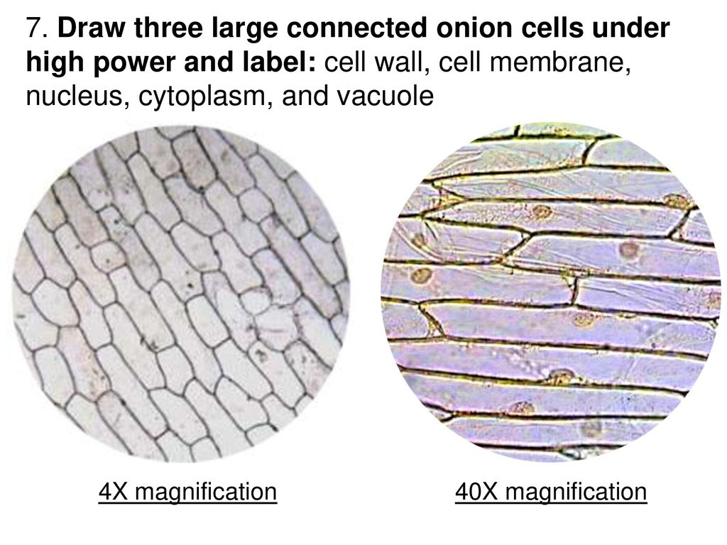
DOC Plant and Animal Cells Microscope Lab - hillsboro.k12.oh.us Make a drawing of one onion cell, labeling all of its parts as you observe them. (At minimum you should observe the nucleus, cell wall, and cytoplasm.) Cheek cells 1. To view cheek cells, gently scrape the inside lining of your cheek with a toothpick. DO NOT GOUGE THE INSIDE OF YOUR CHEEK! (We will observe blood cells in a future lab!!) 2.
Onion Root Tip Mitosis - Stages, Experiment and Results - MicroscopeMaster · Place a cap/lid onto the vial (ensure that the cap/lid has a pinprick hole) and place the vial in the water bath (at 55 degrees C) for about 5 minutes - This enhances the staining process · Using the forceps, remove the root tips from the vial of stain and place them onto a clean microscope glass slide
Microscopy Practical (Onion Cells) | Teaching Resources Presentation and practical handout for observing onion cells under a light microscope for teaching and revision. A step by step visual guide for all abilities. Can be used as a distance based learning tool during local covid lockdown and in classes where practicals are on-hold due to coronavirus. Content covered: Light microscope parts
Vacuole Function and Structure - Extra Space Storage - Rs ... How to see the vacuole under a microscope. Although the vacuole does not take as much dye as other organelles of the cell (the vacuole does not contain many stainable constituents), you can still see and study the structure of vacuoles under a compound microscope. The trick is to use dyes that can stain the cell sap inside the vacuole.
onion cells under a microscope labeled Onion epidermal cells, iodine stain, 400X. distichum 'Spence' (C and D), and sorghum cv BTx623 (E and F) grown in freshwater were imaged under a light microscope Location Tiruvanmiyur, Chennai 600041 Financial Sentiment Analysis Github Label an de diagram of a stomatal apparatus 2 four parts on the diagram Inference The cells observed under the ...
Onion Root Mitosis - Microscopy-UK Onions have larger chromosomes than most plants and stain dark. The chromosomes are easily observed through a compound light microscope. The cells pictured below are located in the apical meristem of the onion root. The apical meristem is an area of a plant where cell division takes place at a rapid rate. Phases of plant cells division:
Onion Cells Under Microscope With Labels - Realtec Find and download Onion Cells Under Microscope With Labels image, wallpaper and background for your Iphone, Android or PC Desktop. Realtec have about 34 image published on this page. onion microscope under cells cepa allium slide footage shutterstock background royalty Pin It Share Download
The Biology Project The Biology Project, an interactive online resource for learning biology developed at The University of Arizona. The Biology Project is fun, richly illustrated, and tested on 1000s of students. It has been designed for biology students at the college and high school level, but is useful for medical students, physicians, science writers, and all types of interested people.
Onion cells microscope Stock Photos and Images - Alamy RM2AM97C0-Onion skin cells under the microscope, horizontal field of view is about 0.61 mm RFHWA476-Onion epidermis with large cells under light microscope. Clear epidermal cells of an onion, Allium cepa, in a single layer. RM2DF6FFJ-Onion epidermis (Allium cepa) showing cells and nucleus. Optical microscope X200.
How to Observe Onion Cells under a Microscope - Blog, She Wrote Place the Onion Peel onto the Slide - You'll want to smooth out any wrinkles with forceps or the end of your pipette Put One Drop or Two of Iodine - onto the top of the onion cell. If you are using Methylene blue, you'll need to apply the stain next to the cover slip after it is down. Go light because too much will mean you can't see the cell well.
Observing Cork Cells Under The Microscope » Microscope Club Place the cork dust on the microscope slide with a drop of water, then add another water droplet on top of the cork sample. Cover the prepared slide with a cover slip. Method 2 Alternatively, slice thin cork slices, making sure that ample light can pass through the slice, allowing you to see the cell layout and the individual cells.
Plant Cell Under Microscope Labeled 40X / Lab Comparing Plant And ... Draw a few water plant cells below, labeling the nucleus, cytoplasm, . Review procedure for this lab . This method allows students to view plant cells under the microscope. Observing onion cells under the microscope. It also depends on plant age and conditions of plant growth, . This method allows students to view plant cells under the microscope.
Onion cells under the microscope: 40X - 100X - 400X - YouTube under the #microscope: 40X - 100X - 400X
Onion Peels Observed Under the Microscope | Confirmation Point Onion Peels Observed Under the Microscope Cells present in onion peel can be observed under microscope. For this onion peels are first isolated. For this experiment outer most scale of the onion is removed and is cut into four equal halves. It is a monocot plant. Then with the help of a pairs of forcep the scale of onion is peeled out.
Microscope Cell Lab: Cheek, Onion, Zebrina | SchoolWorkHelper The onion epidermis cell is the only cell that has a cell wall. In addition, it is the only cell that has a chloroplast, where photosynthesis can happen. The cheek epithelium cell is the only one that has centrioles, the barrel-shaped organelle that is responsible for helping organize chromosomes during cell division.
Onion Plant Cell Under Microscope Labeled / Onion Cells - Onion ... Onion Plant Cell Under Microscope Labeled / Onion Cells - Onion epidermis with pigmented large cells.. Under the microscope, animal cells appear different based on the type of the cell. Human cheek cells and to record observations and draw their labelled diagrams. However, it is too small to see through a microscope.
Onion Cells Stock Photos, Pictures & Royalty-Free Images - iStock Onion epidermis with large cells under microscope Onion epidermis under light microscope. Purple colored, large epidermal cells of an onion, Allium cepa, in a single layer. Each cell with wall, membrane, cytoplasm, nucleus and large vacuole. Photo. Microscope image of plant cells with three nuclei in anaphase
What organelles are in an onion cell? - Biology Stack Exchange You cannot see most of these as they appear translucent as well as being too small to see under the light microscope. You need an electron microscope to view these. ... chloroplasts are not present in an onion cell as it is not a photosynthesising cell. This is a typical onion cell slide with labels: Share. Improve this answer. Follow edited ...
Lesson 3: Onion Dissection & "Look at the Plant Cells" Preparing onion cells slide for a microscope Peel the brown skin away from the outside of the onion. Take one layer of the onion flesh and carefully cut out a piece. On the inside of this piece is a thin sheet of the membrane. Use tweezers or dissection needle to peel the membrane away.










Post a Comment for "43 onion cells under microscope with labels"