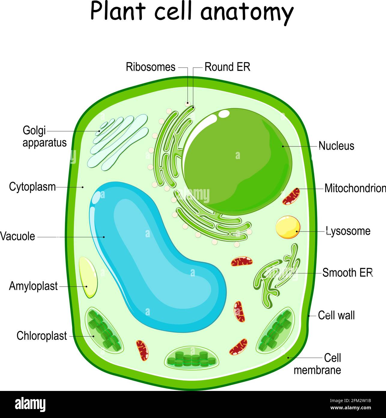
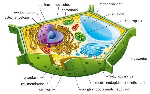
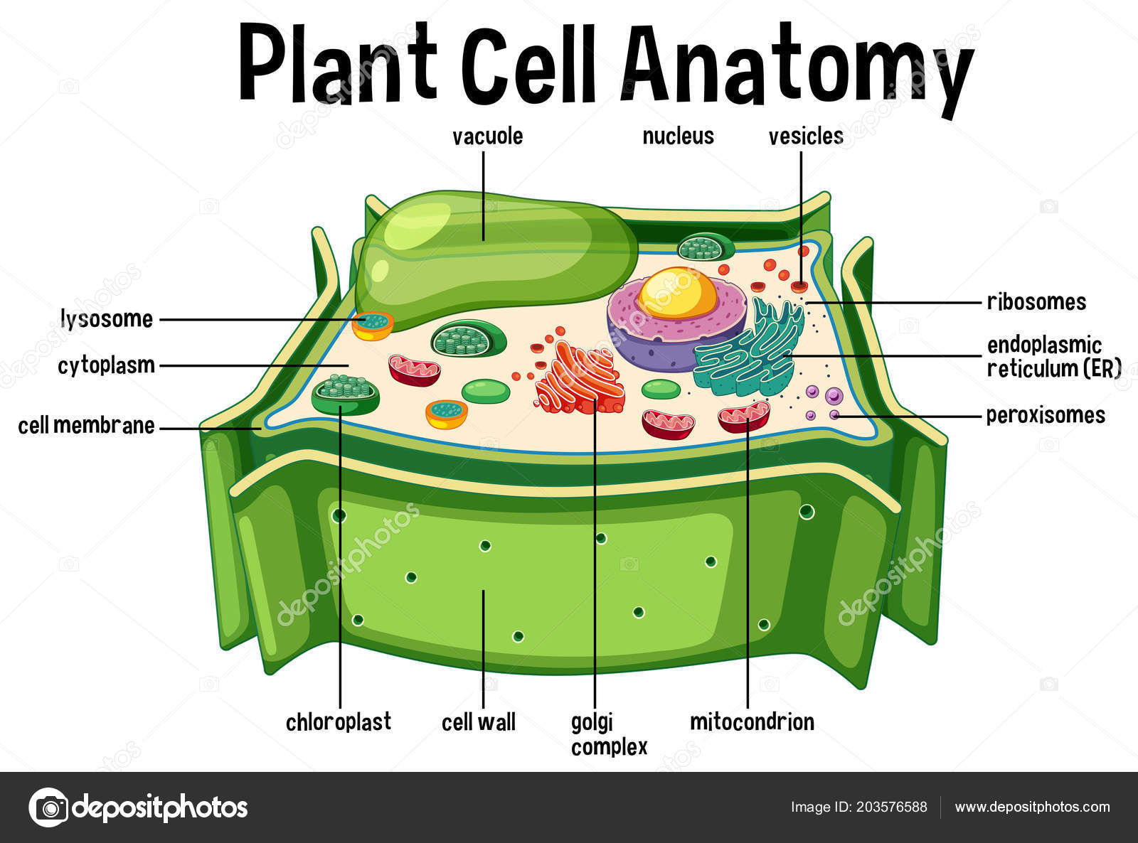
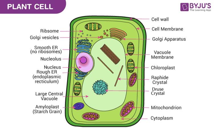
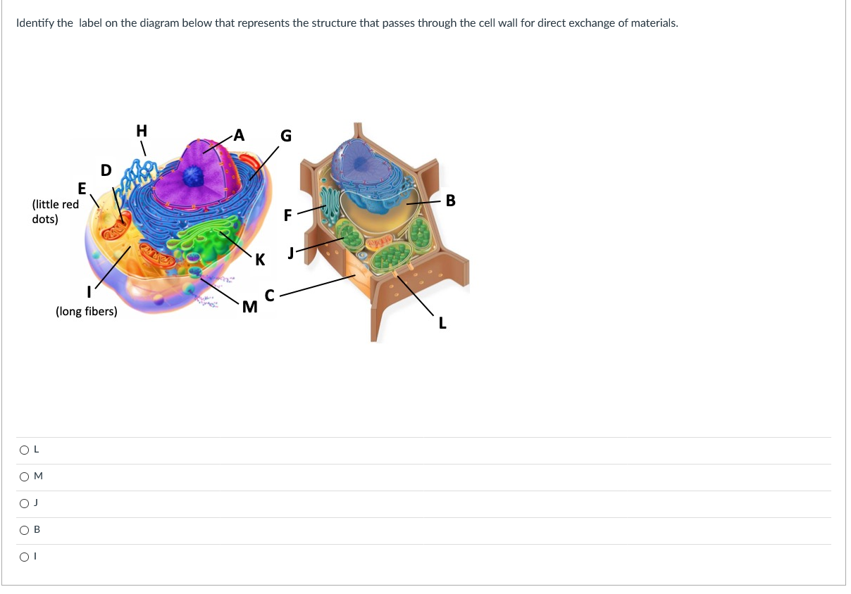
44 cell wall diagram with labels
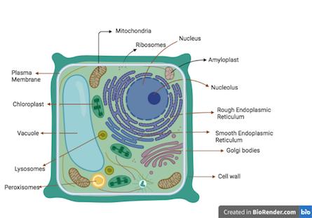
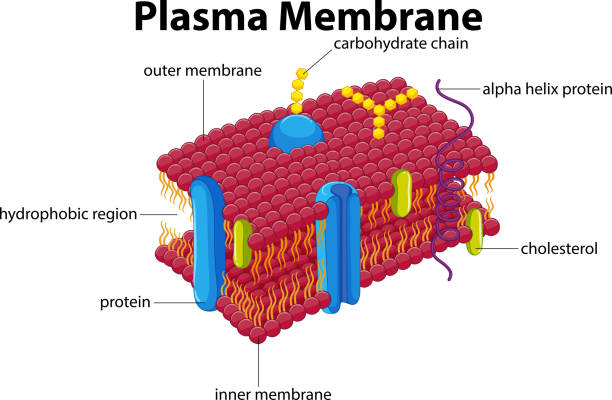
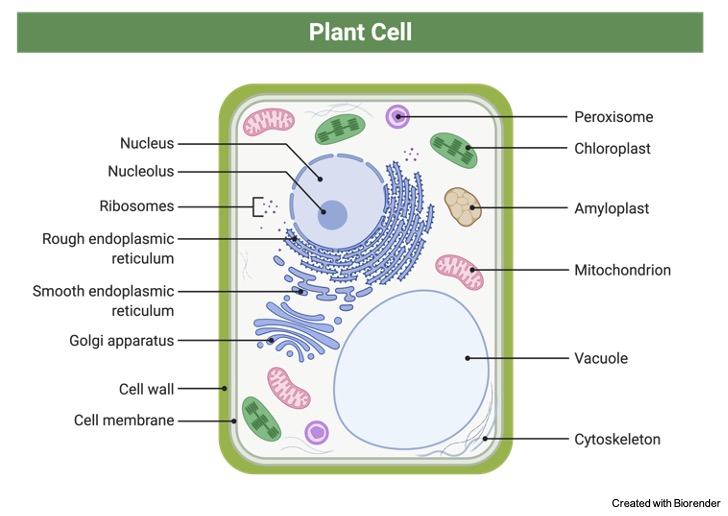
Plant Cell- Definition, Structure, Parts, Functions, Labeled Diagram Functions of the plant cell (plasma) membrane. In-plant cells the cell membrane separated the cytoplasm from the cell wall. It has a selective permeability hence it regulates the contents that move in and out of the cell. It also protects the cell from external damage and provides support and stability to the cell. Protein Targeting (With Diagram) | Molecular Biology ADVERTISEMENTS: Let us make an in-depth study of the protein targeting. After reading this article you will learn about: 1. Introduction to Protein Targeting 2. Signal Sequence 3. Transport of Proteins into ER 4. Signal Sequence Recognition Mechanism 5. Role of Golgi Complex in Protein Transportation 6. Transport of Proteins from Golgi to Lysosomes 7. […]
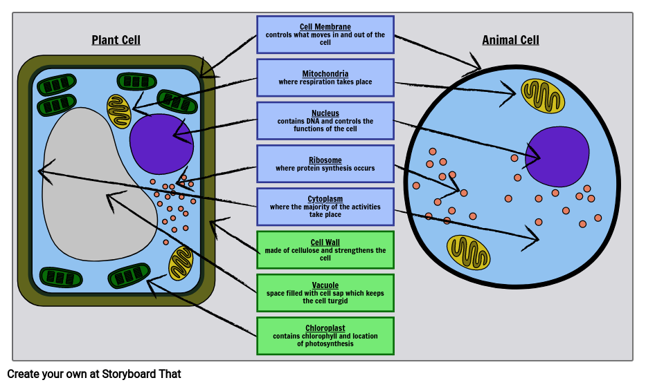
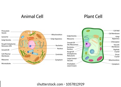
Plant and Animal Cell: Labeled Diagram, Structure, Function - Embibe Cell Wall: 1. Non-living, rigid, outer boundary. 2. Made up of cellulose, hemicellulose, pectin, lignin, etc. 3. There are many layers, like the middle layer, primary cell wall in a typical plant cell wall. 4. Fungal cell wall is made up of chitin (not cellulose). 5. Protective and provide shape and size. 6. Found only in plant cells. Plasma ...
Cell wall diagram with labels
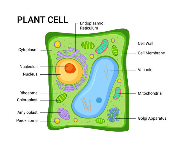
Human Cell Diagram, Parts, Pictures, Structure and Functions Diagram of the human cell illustrating the different parts of the cell. Cell Membrane. The cell membrane is the outer coating of the cell and contains the cytoplasm, substances within it and the organelle. It is a double-layered membrane composed of proteins and lipids. The lipid molecules on the outer and inner part (lipid bilayer) allow it to ... Structure of Bacterial Cell (With Diagram) - Biology Discussion Cell wall: It is a tough and rigid structure of peptidoglycan with accessory specific materials (e.g. LPS, teichoic acid etc.) surrounding the bacterium like a shell and lies external to the cytoplasmic membrane. It is 10-25 nm in thickness. It gives shape to the cell. Nucleus: The single circular double-stranded chromosome is the bacterial genome. Labeled Plant Cell With Diagrams | Science Trends The parts of a plant cell include the cell wall, the cell membrane, the cytoskeleton or cytoplasm, the nucleus, the Golgi body, the mitochondria, the peroxisome's, the vacuoles, ribosomes, and the endoplasmic reticulum. Parts Of A Plant Cell The Cell Wall Let's start from the outside and work our way inwards.
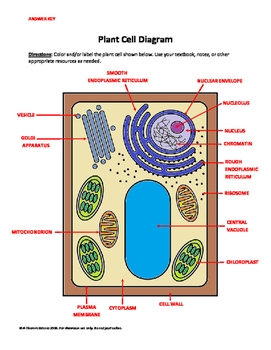
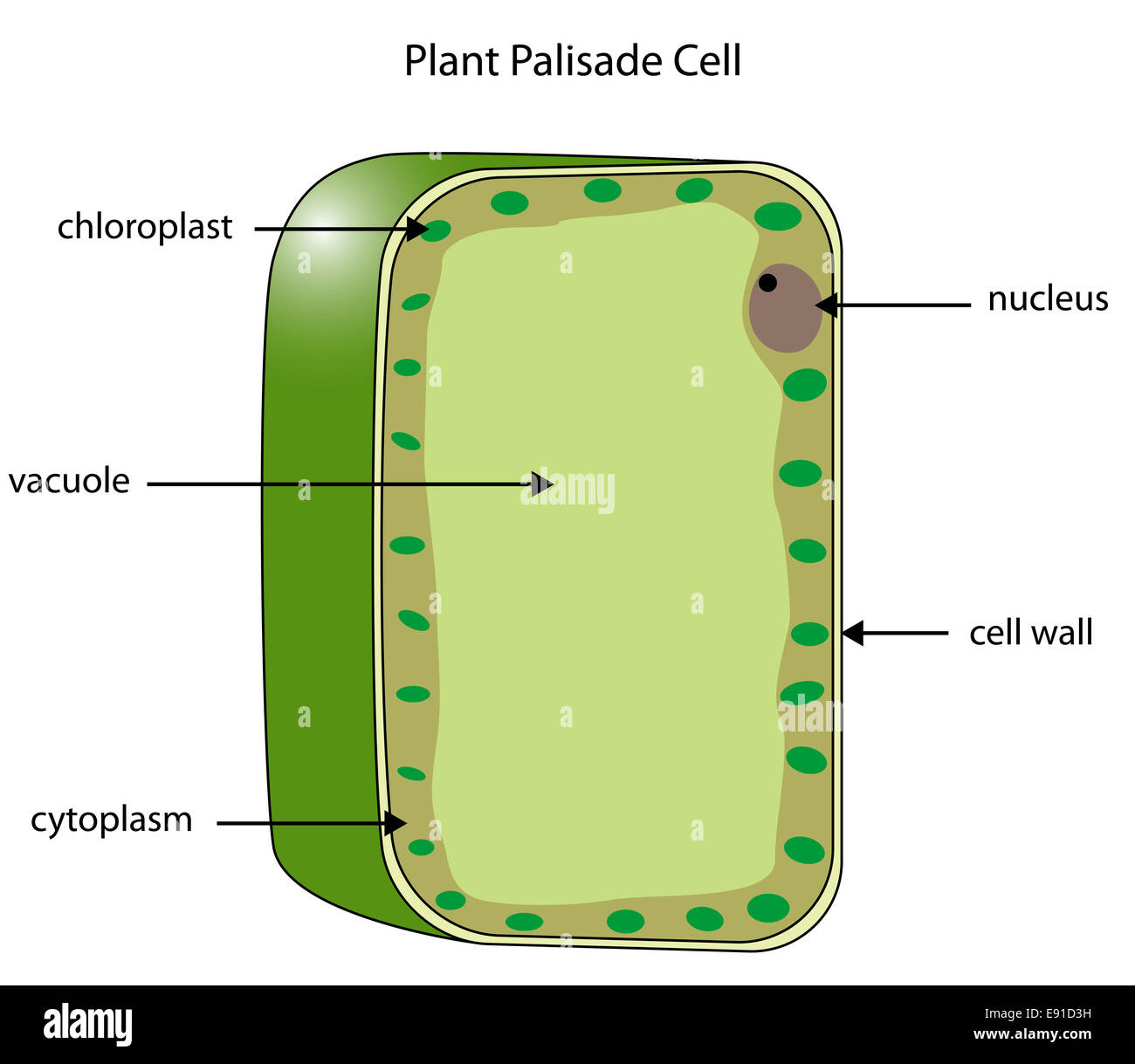
Cell wall diagram with labels. The Cell - ScienceQuiz.net The diagram shows a plant cell as seen under a microscope. Two of the labels are incorrect. What are they?? Vacuole and chloroplast? Vacuole and cytoplasm? Nucleus and chloroplast? Cell wall and cell membrane; The diagram shows a plant cell. Identify parts A and B. ? A = cell wall and B = nucleus.? A = cell wall and B = cytoplasm.? A = cell membrane and B = nucleus.? A = … Structure of Cell Wall (With Diagram) | Plants - Biology Discussion Cells with secondary wall consist of five layers a three layered secondary wall, the primary wall and the middle lamella. In some cells, such as primary xylem, the secondary thickening materials are laid down in such a way that various patterns are formed on the cell wall, e.g. annular, spiral, reticulate, scalariform and pitted. Origin 2018 Feature Highlights Fixed cell referencing is supported Use script-accessible functions and/or custom expressions Results update when source cells change, or with other changes such as data filter Assign formula to User-defined Parameter Rows to easily set up calculations such as calculating mean and std. deviation as in this example. Enter formula in one cell and drag-and-extend to the right … Free Cell Diagram Software with Free Templates - EdrawMax - Edrawsoft A plant cell diagram has a cell wall, cell membrane, nucleus, organelles, endoplasmic reticulum, ribosome, plastids, mitochondria, and multiple vesicles. Animal Cell Diagram An animal cell diagram describes a cell structure enclosed by a plasma member, and it has a nucleus with a membrane and organelles. Neuron Diagram
Structure of Fungal Cell (With Diagram) | Fungi - Biology Discussion The basic structural constituent of the cell wall in the Zygomycetes and higher fungi (Ascomycetes and Basidiomycetes) is chitin. It is a polysaccharide based on the nitrogen containing sugar (glucosamine). It is probable that more or less closely associated with chitin in the cell wall are pectic materials, protein, lipids, cellulose, callose ... A Labeled Diagram of the Plant Cell and Functions of its Organelles The cell membrane is a thin layer made up of proteins, lipids, and fats. It forms a protective wall around the organelles contained within the cell. It is selectively permeable and thus, regulates the transportation of materials needed for the survival of the organelles of the cell. Function: Protects the cell from its surroundings. Structure of Cell: Definition, Types, Diagram, Functions - Embibe The cell wall lies outside the plasma membrane. The plant cell wall is the outermost layer of a plant cell. It is mainly composed of cellulose which is a complex substance and provides structural strength to plants or cells. Cell walls permit the cells of plants to withstand very dilute (hypotonic) external media without bursting. Cell walls ... Cell Organelles- Definition, Structure, Functions, Diagram - Microbe Notes In a plant cell, the cell wall is made up of cellulose, hemicellulose, and proteins while in a fungal cell, it is composed of chitin. A cell wall is multilayered with a middle lamina, a primary cell wall, and a secondary cell wall. The middle lamina contains polysaccharides that provide adhesion and allow binding of the cells to one another.
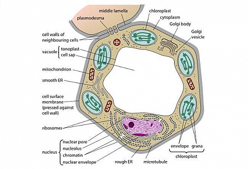
Cell Diagram To Label Teaching Resources | Teachers Pay Teachers Interactive Google Slides activities to help your students learn about cell organelles! Custom clip art is used to clearly illustrate each organelle in a diagram for them to label and then describe the structure and function of. With an open response and a scaffolded fill-in-the-blanks version, differentiation is simple! Plant Cell Wall- Definition, Structure, Functions, Diagram Structure of Plant Cell wall It is derived from the living protoplast. It consists of the middle lamella, primary cell wall, plasmodesmata, secondary cell wall, and pits. Middle lamella After the cytokinesis, it is the first-formed layer. It is present in between the two adjacent cells. It is made up of calcium and magnesium pectate. Nervous System – Medical Terminology for Healthcare Professions Neurons are the primary type of cell that most anyone associates with the nervous system. They are responsible for the computation and communication that the nervous system provides. They are electrically active and release chemical signals to target cells. Glial cells, or glia, are known to play a supporting role for nervous tissue. Ongoing research pursues an expanded role that glial … Cell Biology - Wiki - Scioly.org 2 days ago · Cell membrane: Functions in transport, the movement of substances in and out of the cell, and in energy production (breakdown of large molecules, photosynthesis). Cell wall: Gives structural strength (rigidity) to the cell. Capsule or slime layer: Jelly-like substance which protects the cell wall from environmental damage. Nucleoid
Bacteria in Microbiology - shapes, structure and diagram - Jotscroll The bacteria shapes, structure, and labeled diagrams are discussed below. Table of Contents Sizes Bacteria Shapes Structure of Bacteria cells Components that make up the structures of a bacterial cell Bacterial Cell Envelope Bacterial Capsule or Slime layer Bacteria Cell Wall Functions of the bacteria cell wall Types of Bacteria Cell walls
Interactive Bacteria Cell Model - CELLS alive Periplasmic Space: This cellular compartment is found only in those bacteria that have both an outer membrane and plasma membrane (e.g. Gram negative bacteria).In the space are enzymes and other proteins that help digest and move nutrients into the cell. Cell Wall: Composed of peptidoglycan (polysaccharides + protein), the cell wall maintains the overall shape of a …
Bacterial Cell Structure Labeling Diagram | Quizlet Cell Wall A semirigid casing that provides structural support and shape for the cell Cytoskeleton Long fibers of proteins that encircle the cell just inside the cytoplasmic membrane and contribute to the shape of the cell Pilus Appendage used for drawing another bacterium close in order to transfer DNA Glycocalyx
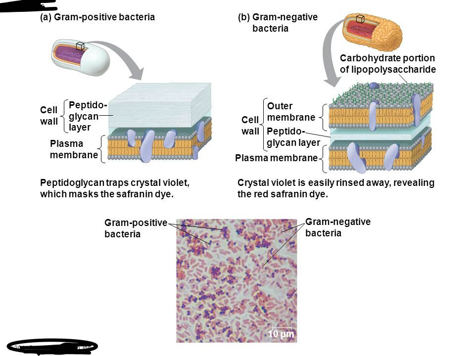
Solved In the following image, the diagram labeled "A" | Chegg.com Explanation :Above statement is false because of gram -( negative ) bacteria cell wall which is protected by su… View the full answer Transcribed image text : In the following image, the diagram labeled "A" represents a gram-negative bacterial cell wall, and the diagram labeled "B" represents a gram-positive bacterial cell wall.
PDF Human Cell Diagram, Parts, Pictures, Structure and Functions Human Cell Diagram, Parts, Pictures, Structure and Functions The cell is the basic functional in a human meaning that it is a self-contained and fully operational living entity. Humans are multicellular organisms with various different types of cells that work together to sustain life. Other non-cellular components in the body include water ...
Cell Wall: Definition, Structure & Function (with Diagram) In general, fungi with cell walls have three layers: chitin, glucans and proteins. As the innermost layer, chitin is fibrous and made up of polysaccharides. It helps make the fungi cell walls rigid and strong. Next, there is a layer of glucans, which are glucose polymers, crosslinking with chitin.
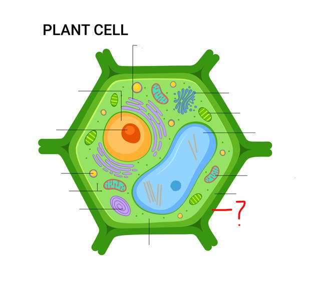
Plant Cell: Diagram, Types and Functions - Embibe Exams Plant Cell Wall It is a rigid layer that is composed of cellulose, glycoproteins, lignin, pectin and hemicellulose. It is located outside the cell membrane and is completely permeable. The primary function of a plant cell wall is to protect the cell against mechanical stress and to provide a definite form and structure to the cell.
History of cell membrane theory - Wikipedia Cell theory has its origins in seventeenth century microscopy observations, but it was nearly two hundred years before a complete cell membrane theory was developed to explain what separates cells from the outside world. By the 19th century it was accepted that some form of semi-permeable barrier must exist around a cell. Studies of the action of anesthetic molecules led to …
03 Label the Cell Diagram | Quizlet Start studying 03 Label the Cell. Learn vocabulary, terms, and more with flashcards, games, and other study tools. ... cell diagram. 18 terms. lugo_janet. Sets found in the same folder. 03 Organelle Functions. 14 terms. muskopf1. 07 Cell Labeling. 11 terms. muskopf1. 03 Cell Transport. 12 terms.
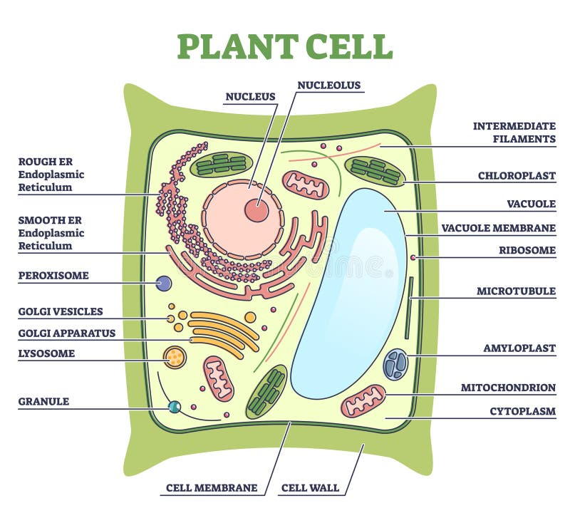
Labeled Plant Cell With Diagrams | Science Trends The parts of a plant cell include the cell wall, the cell membrane, the cytoskeleton or cytoplasm, the nucleus, the Golgi body, the mitochondria, the peroxisome's, the vacuoles, ribosomes, and the endoplasmic reticulum. Parts Of A Plant Cell The Cell Wall Let's start from the outside and work our way inwards.
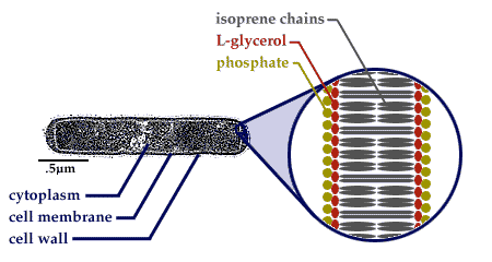
Structure of Bacterial Cell (With Diagram) - Biology Discussion Cell wall: It is a tough and rigid structure of peptidoglycan with accessory specific materials (e.g. LPS, teichoic acid etc.) surrounding the bacterium like a shell and lies external to the cytoplasmic membrane. It is 10-25 nm in thickness. It gives shape to the cell. Nucleus: The single circular double-stranded chromosome is the bacterial genome.
Human Cell Diagram, Parts, Pictures, Structure and Functions Diagram of the human cell illustrating the different parts of the cell. Cell Membrane. The cell membrane is the outer coating of the cell and contains the cytoplasm, substances within it and the organelle. It is a double-layered membrane composed of proteins and lipids. The lipid molecules on the outer and inner part (lipid bilayer) allow it to ...

:max_bytes(150000):strip_icc()/cell-membrane-373364_final-5b5f300546e0fb008271ce52.png)







:background_color(FFFFFF):format(jpeg)/images/article/en/cellular-organelles/YPFQDP4aMP9RH4TYp85YQ_Cell_membrane_V2.png)









Post a Comment for "44 cell wall diagram with labels"