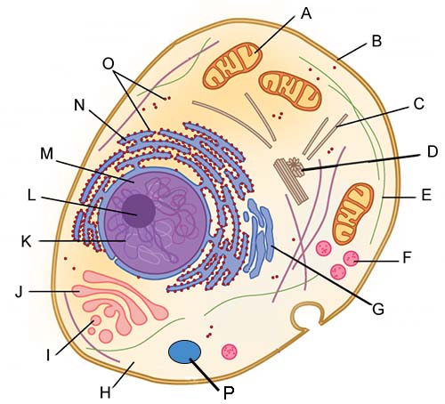
42 cell image with labels
2,284 Animal cell labeled Images, Stock Photos & Vectors - Shutterstock Find Animal cell labeled stock images in HD and millions of other royalty-free stock photos, illustrations and vectors in the Shutterstock collection. Thousands of new, high-quality pictures added every day. Plant Cell Label Images, Stock Photos & Vectors | Shutterstock Find plant cell label stock images in HD and millions of other royalty-free stock photos, illustrations and vectors in the Shutterstock collection. Thousands of new, high-quality pictures added every day.
en.wikipedia.org › wiki › Flow_cytometryFlow cytometry - Wikipedia Labels, dyes, and stains can be used for multi-parametric analysis (understand more properties about a cell). Immunophenotyping is the analysis of heterogeneous populations of cells using labeled antibodies [35] and other fluorophore containing reagents such as dyes and stains.

Cell image with labels
Plot Images and Labels | Kaggle Plot Images and Labels Python · NIH Chest X-rays. Plot Images and Labels. Notebook. Data. Logs. Comments (0) Run. 175.6s - GPU. history Version 3 of 3. GPU Data Visualization Image Data. Cell link copied. License. This Notebook has been released under the Apache 2.0 open source license. Continue exploring. Data. 1 input and 0 output. arrow ... Real Cell Gallery - University of Utah Image Credits. Breast Cancer Cells: National Cancer Institute. Diatom: Robert Pickett. Haloquadratum walsbyi: Mike Dyall-Smith. Heart Muscle Cell: Jose Martin and Jose Enrique Muzquiz. Influenza A: Frederick Murphy, CDC Public Health Image Library. Mold hyphae: Y. Tambe; licensed under CC BY-SA 3.0. Cropped image and added labels. Osteocyte ... How to add images to labels in Google Docs? 5. Add images and text. Import your image or logo inside the label. Go to the "Insert" menu at the top, then select "Image" and "Upload from computer". You can also drag and drop your image from your computer inside the first cell. Once you insert your image, add the text that will be displayed on your label.
Cell image with labels. › figureguidelinesDigital image guidelines: Cell Press Captions and atom labels* = Arial, 7 pt; Atom labels "Show labels on Terminal Carbons" and "Hide Implicit Hydrogens" should be unchecked. For initial submission, ChemDraw files must either be embedded in the text or supplied separately as TIFF or PDF files. For final submission, please use our one-column or full-page template. › cell › fulltextIdentifying Medical Diagnoses and Treatable Diseases ... - Cell Feb 22, 2018 · Before training, each image went through a tiered grading system consisting of multiple layers of trained graders of increasing expertise for verification and correction of image labels. Each image imported into the database started with a label matching the most recent diagnosis of the patient. Label Cell Parts | Plant & Animal Cell Activity | StoryboardThat Click "Start Assignment". Find diagrams of a plant and an animal cell in the Science tab. Using arrows and Textables, label each part of the cell and describe its function. Color the text boxes to group them into organelles found in only animal cells, organelles found in only plant cells, and organelles found in both cell types. › food › food-labeling-nutritionChanges to the Nutrition Facts Label | FDA Mar 07, 2022 · Manufacturers with $10 million or more in annual sales were required to update their labels by January 1, 2020; manufacturers with less than $10 million in annual food sales were required to ...
Plant Cell Images With Labels - Extra Cedit Label The Plant And Animal ... Pictures cells that have structures unlabled, students must write the labels in, . 4000+ vectors, stock photos & psd files. A typical plant cell organelles include cell wall, cell membrane, cytoskeleton, . free for commercial use high quality images. A cell through creating, labeling, writing, and presenting to the class. Animal Cell Labeled Pictures, Images and Stock Photos Browse 116 animal cell labeled stock photos and images available, or start a new search to explore more stock photos and images. Components of Eukaryotic cell, nucleus and organelles and plasma... Labelled diagrams of typical animal and plant cells with editable layers. Golgi apparatus. Animal Cell Labeled Diagram Pictures, Images and Stock Photos Browse 19 animal cell labeled diagram stock photos and images available, or start a new search to explore more stock photos and images. Newest results. Diagrams of animal and plant cells. Labelled diagrams of typical animal and plant cells with editable layers. Golgi apparatus or Golgi body. Blood Cell Images | Kaggle This dataset contains 12,500 augmented images of blood cells (JPEG) with accompanying cell type labels (CSV). There are approximately 3,000 images for each of 4 different cell types grouped into 4 different folders (according to cell type). The cell types are Eosinophil, Lymphocyte, Monocyte, and Neutrophil.
Image Data Labelling and Annotation — Everything you need to know Labeled bottle of blueberries (Photo by Debby Hudson on Unsplash). Data labelling is an essential step in a supervised machine learning task. Garbage In Garbage Out is a phrase commonly used in the machine learning community, which means that the quality of the training data determines the quality of the model. The same is true for annotations used for data labelling. Label-free imaging of live cells | CytoSMART Cells can be highly dynamic with changes in their morphology and behavior. Live-cell label-free imaging enables insight into these changes over time. Label-free imaging enables the identification and quantification of cellular events such as cell division, proliferation, motility, migration, differentiation, and death (Figure 4). These are fundamental processes in development, tissue repair, and immune regulation. Labelling cell quiz - Teaching resources - Wordwall Animal Cell Labelling Labelled diagram. by Claudine11. Labelling a Plant Cell Labelled diagram. by Hpaterson2. High school KS3 Biology Science. Plant Cell Labelling Challenge Labelled diagram. by Swright. KS4. Yeast Cell: Labelling Labelled diagram. How to Label Images for Object Detection, Step by Step How to Use this tool. Click on "Open Dir" and select the folder where you have saved your images that you need to label. Then click on "Change Save Dir" here, you need to select the directory to save your label file. This directory should be different from the image directory. Now you can use "Create Rectbox" to draw boxes over the ...
Animal Cell - Free printable to label + Color -kidCourses.com Can you label and color these important parts of the animal cell?. NUCLEUS control center for cell (cell growth, cell metabolism, cell reproduction). NUCLEOLUS synthesizes rRNA. RIBOSOMES the site of protein building, this is where translation takes place (mRNA in language of nucleic acids is translated into the language of amino acids). RER (Rough Endoplasmic Reticulum) synthesizes proteins ...
Plant and Animal Cells - Labeled Graphics A compilation of plant and animal cell images with organelles and major structures labeled. Students can print images to help them learn the cell. Other Cell Resources. Cheek Cell Lab - observe cheek cells ... Cell Model - create a cell from household and kitchen items, rubric included Cell Research & Design - research cells on the web, ...
› site-mapAutoblog Sitemap Here's how to disable adblocking on our site. Click on the icon for your Adblocker in your browser. A drop down menu will appear. Select the option to run ads for autoblog.com, by clicking either ...
Add graphics to labels - support.microsoft.com Insert a graphic and then select it. Go to Picture Format > Text Wrapping, and select Square. Select X to close. Drag the image into position within the label. and type your text. Save or print your label. Note: To create a full sheet of labels, from your sheet with a single label, go to Mailings > Labels and select New Document again. This ...
Labeled Plant Cell With Diagrams | Science Trends Plant cells contain many organelles such as ribosomes, the nucleus, the plasma membrane, the cell wall, mitochondria, and chloroplasts. In addition, plant cells differ from animal cells in a number of key ways. Examining a diagram of the plant cell will help make the differences clearer. Let's go over the individual components of plant cells ...
› en-us › autosUsed cars and new cars for sale – Microsoft Start Autos - MSN Find new and used cars for sale on Microsoft Start Autos. Get a great deal on a great car, and all the information you need to make a smart purchase.
Cell Cycle Labeling | Cell cycle, Biology worksheet, Mitosis Students label the image of a cell undergoing mitosis and answer questions about the cell cycle: interphase, prophase, metaphase, anaphase, and telophase. Find this Pin and more on Education by Indra Sadiq. Biology Lessons Teaching Biology Science Lessons Science Activities Cell Biology Science Biology Science Education Life Science Tissue Biology
Automated Image Processing Workflow for Morphological Analysis of ... The workflow developed in this work is implemented in the python programming language using the open-source NumPy, SciPy28 and scikit-image packages.29 It first performs image segmentation to generate labels for each pixel in the image, where each label is uniquely associated with a cell in the image. The labeled images are then used to extract the statistics of various cell morphology measures of interest.
In Silico Labeling: Predicting Fluorescent Labels in Unlabeled Images: Cell The unlabeled image that is the basis for the prediction and the images of the true and predicted fluorescent labels are organized similarly to Figure 4, but in the first row the true and predicted nuclear (DAPI) labels have been added to the true and predicted images in blue for visual context, and in the second row the true and predicted neuron (TuJ1) labels were added. Outset 1 shows a false positive, in which a neuron was wrongly predicted to be a motor neuron.
Bacteria Cell Structures with Labels Stock Vector - Dreamstime Bacteria Cell Structures with labels. Royalty-Free Vector. Bacterial cell structures labeled on a bacillus cell with nucleoid DNA and ribosomes. External structures include the capsule, pili, and flagellum. Morphology of internal structures of bacteria. cell anatomy bacteria,
All About Cells & DNA - StartsAtEight | Cells project, Animal cell ... Feb 1, 2015 - A labeled diagram of an animal cell, and a glossary of animal cell terms. Learn about the different parts of a cell.
Cell Labeling (Remote) - The Biology Corner The format is Google Slides, where the cell images were placed as slide backgrounds, that way only the words can be manipulated by the user. The first set gives students word boxes to drag and drop into the correct location. Boxes have labels for the various organelles, like mitochondria, ribosomes, lysosomes, endoplasmic reticulum, and nucleus.
A weakly supervised deep learning approach for label-free ... - cell.com The model was trained on cell images with individual labels. We defined a naive annotation, where all the cells from the same patient specimen were assigned with the respective patient's health status (i.e., healthy or diseased). The model was trained on the basis that SS patients have a larger percentage of morphologically atypical cells.
Labeled Cell - an overview | ScienceDirect Topics Red blood cells labeled with 99m Tc are used to image gastrointestinal bleeding. For this, dynamic planar images with a frame rate of 1 frame per 60 s are used to identify the bleeding source. Planar imaging is also frequently used for ventilation-perfusion (V/Q) scan of lungs to investigate pulmonary embolism using 133 Xe gas or 99m Tc-DTPA aerosols.
› articles › s41587/021/01094-0Whole-cell segmentation of tissue images with human-level ... Nov 18, 2021 · We generated whole-cell segmentation labels for 15 of these images manually using HH3 to define the nucleus and CD3, CD14, CD56, HLAG and vimentin to define the shape of the cells in the image.
Animal Cell Diagram with Label and Explanation: Cell ... - Collegedunia Animal cell is a typical Eukaryotic cell enclosed by a plasma membrane containing nucleus and organelles which lack cell walls, unlike all other Eukaryotic cells. The typical cell ranges in size between 1-100 micrometers. The lack of cell walls enabled the animal cells to develop a greater diversity of cell types.
Structure of Bacterial Cell (With Diagram) - Biology Discussion Capsule: It is an outer covering of thin jelly-like material (0.2 μm in width) that surrounds the cell wall. Only some bacterial species possess capsule. Capsule is usually made of polysaccharide (e.g. pneumococcus), occasionally polypeptide (e.g. anthrax bacilli) and hyaluronic acid (e.g. streptococcus).


Post a Comment for "42 cell image with labels"